Anatomy of the Heel: Understanding the Complex Structure

Introduction

The heel is more than just the back part of your foot; it’s a complex structure comprising various bones, muscles, ligaments, and tendons. Understanding the anatomy of the heel can provide valuable insights into conditions like heel painplantar fasciitis, and Achilles tendinitis. This article will guide you through the essential components of the heel’s anatomy.

The Calcaneus: The Foundation of the Heel

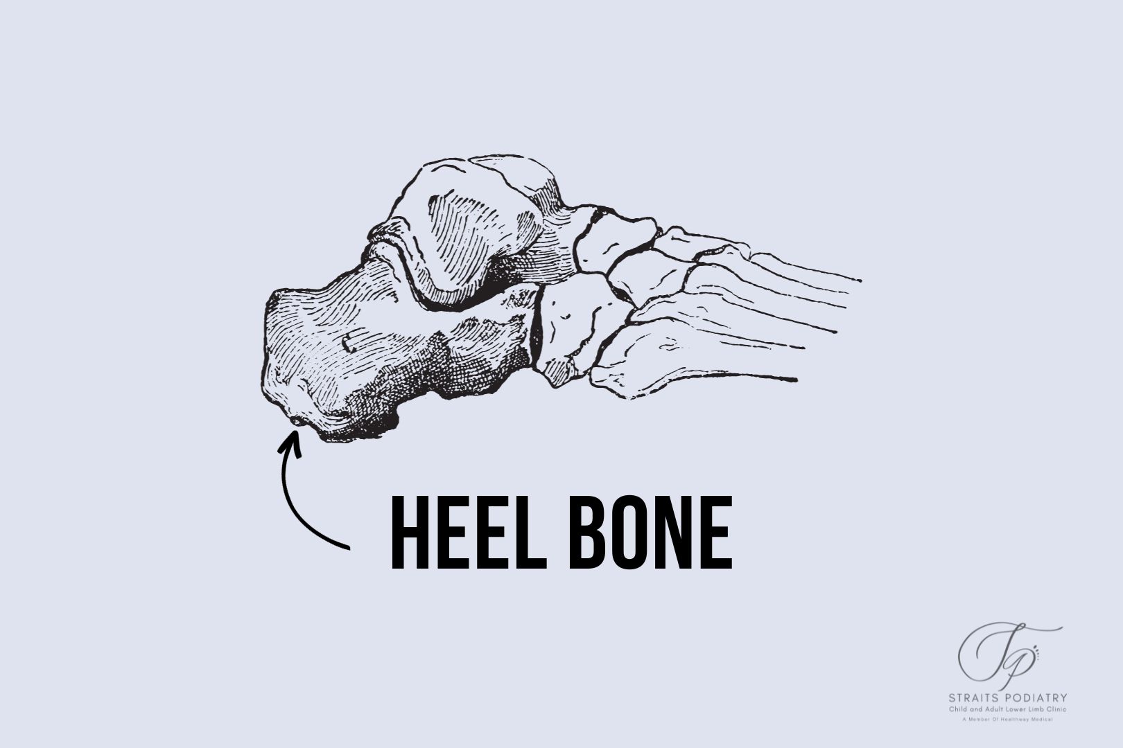
The calcaneus, or heel bone, is the largest bone in the foot. It serves as the foundation for the rear part of the foot and plays a crucial role in walking and running by providing a lever for muscles to exert force. It also bears most of our body load when walking or running, taking high impact and pressure every step we take.

Ligaments and Tendons

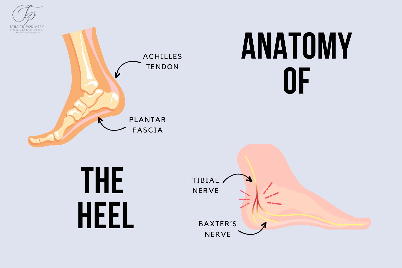
There are two main ligamentous and tendinous structure that attaches to the heel bone, and they play a key role in the function and stability of the foot:

  • Plantar Fascia: This thick fibrous band of tissue connects the bottom of the heel bone to the toes and supports the arch of the foot.
  • Achilles Tendon: This tendon connects the calf muscles to the back of the heel bone and is essential for running, jumping, and standing on the toes.

Muscles Involved in Heel Function

Several intrinsic muscles originate from the region of the heel. They assist the larger tendons in the functioning of the foot:

  • Quadratus Plantae: Helps to assist the flexor digitorium longus in flexing the lesser toes
  • Flexor Digitorum Brevis: This is the secondary muscle that helps flex the lesser toes and is also thought to provide arch support.
  • Abductor Hallucis: One of the larger muscles within the foot, located at the arch area. This helps to abduct and flex the big toe and provide foot stability.
  • Abductor Digiti Minimi: Helps to abduct and flex the little toe. A muscle that not every individual can isolate its contraction and movement.

Soft Tissues and Nerves

The soft tissues and nerves within the heel plays an important part in cushioning the impact and providing sensory-motor control respectively. The key structures are:

  • Fat Pad: A beautiful honeycomb structure containing fatty tissue that sits under the heel bone. It helps to absorb the impact of walking and running.
  • Tibial Nerve: The largest nerve going into the foot. This nerve runs from the inner side of the ankle to under the heel and can be involved in conditions like tarsal tunnel syndrome.
  • Baxter’s Nerve: A nerve branch originating from the lateral plantar nerve. Compression of this nerve causes Baxter’s nerve entrapment.

How Anatomy Affects Heel Pain

Understanding the anatomy of the heel can help medical professionals diagnose and treat heel pain more effectively. For example, inflammation of the plantar fascia leads to plantar fasciitis, while issues with the Achilles tendon can result in Achilles tendinitis. It is also crucial for medical professional to know the anatomy well to administer treatment to the correct structure.

Conclusion

The heel is a complex structure with various components working in harmony to enable movement and provide support and stability. Understanding its anatomy can offer valuable insights into the causes and treatments of heel-related conditions.

Related Articles

Picture of Jackie Tey

Jackie Tey

Chief Podiatrist, B.Pod(Hons). Your foot and lower limb specialist passionate about raising awareness for foot and lower limb health.

Share this with someone you know

Search

You May Also Like

Do You Have A Question? Ask Us...

Search

Do You Have A Question? Ask Us...

You May Also like

About Us

Conditions

Treatments

Feet

Ankle

Leg

Knee

Paediatric Conditions

Rheumatology

Skin & Nails

Diabetic Foot

Heel Pain

Foot Pain

Plantar Fasciitis

Achilles Tendonitis

Flat Feet

High Arch Foot Type

Arch Pain

Bunions

Hallux Rigidus

Sesamoiditis

Plantar Plate Injury

Metatarsalgia

Foot Drop

Tarsal Tunnel Syndrome

Baxter’s Nerve Entrapment

Clawed and Hammer Toes

Heel Spurs

Morton’s Neuroma

Accessory Navicular

Ankle Injuries

Posterior Tibial Tendon Dysfunction

Chronic Ankle Sprain

Acute Ankle Sprain

Talar Dome Injuries

Ankle Impingement

Knee Pain

Runner’s Knee

Jumper’s Knee

Meniscus Tear

Iliotibial Band Syndrome

Knee Osteoarthritis

Children’s Flat Feet

In-toe Walking

Toe Walking

Knock Knees

Paediatric Heel Pain

Juvenile Bunions

Osteochondrosis

Kohler’s Disease

Iselin’s Disease

Osgood-Schlatter Disease

Rheumatoid Arthritis

Gout

Fungal Nails

Plantar Warts

Ingrown Toenails

Fungal Skin

Corns and Calluses

Diabetic Peripheral Neuropathy

Diabetic Foot Ulcers

Peripheral Arterial Disease

Charcot Foot