Os Navicular Syndrome (Accessory Navicular)

Introduction

Os Navicular Syndrome, also known as accessory navicular, is a condition that causes foot pain and discomfort around the inner arch area. A person suffering from the pain will have difficulty taking long walks or performing sports. If left untreated, it can cause long-term chronic pain and affect mobility. This condition is commonly misdiagnosed as posterior tibial tendonitis due to the similarities in the signs and symptoms. Therefore, in this article, we will list the common causes, signs and symptoms, and the treatment options typically available for Os Navicular Syndrome.

What is Accessory Navicular?

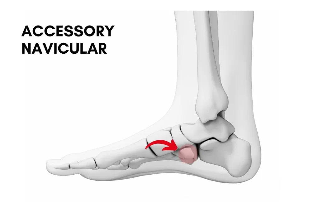
An accessory navicular is a small extra bone or cartilage located near the inner side of the foot, just above the arch, where the navicular bone typically sits. This accessory bone is present in some individuals due to a congenital condition where an additional piece of bone forms during development. While many people with an accessory navicular experience no symptoms, in others, it can lead to Os Navicular Syndrome, where pain and inflammation occur in the area. The condition often occurs when physical activity or specific movements trigger discomfort in the foot.

Need Help? See Our Podiatrist Today

Causes of Os Navicular Syndrome

Os Navicular Syndrome occurs when the accessory navicular bone becomes irritated, inflamed, or painful. The condition is typically a result of overuse by the posterior tibial tendon pulling on the accessory navicular bone. The common causes include:

  • Overuse or Repetitive Stress: High-impact activities such as running, jumping, or intense sports can cause stress to the accessory navicular, leading to inflammation or pain.
  • Foot Deformities: Abnormal foot structure, such as flat feet, further stresses the accessory navicular, leading to pain and discomfort.
  • Trauma or Injury: Direct injury to the foot or a sudden impact can cause trauma to the accessory navicular, resulting in pain and discomfort.
  • Ankle Sprains: Acute injuries like ankle sprains can trigger inflammation of the accessory navicular
  • Tight Footwear: Shoes that don’t provide sufficient room and adequate support can lead to gait abnormality and worsening symptoms.

Causes of Arch Pain

If any of the above symptoms are hitting the spot, it’s time to know why your arch is unhappy. In a concrete jungle like Singapore, most people don’t wear shoes that are cushioned enough, or they overtrain on hard, unforgiving surfaces for their beloved sports. Over time, they start to develop conditions that give rise to the pain in their arch, such as:
  • Plantar Fasciitis: undisputedly one of the most common causes of arch pain. Plantar fasciitis is a result of inflammation of the plantar fascia, a thick band of connective tissue that holds the arch. The pain is often most intense with the first few steps in the morning or after a long period of rest.
  • Posterior Tibial Tendon Dysfunction (PTTD): an overuse injury of a crucial tendon that controls the archWhen this tendon becomes inflamed or weakened, it loses its function, leading to a gradual collapse of the arch. That is why it is also known as adult-acquired flat foot deformity. PTTD often causes pain along the inner side of the arch, which typically worsens after activity.
  • Flexor Hallucis Tendonitis: an injury to the tendon that helps to claw the big toe. Dancers and ballerinas are more prone to this condition because they frequently bend and flex their toes. Flexor hallucis tendonitis tends to cause pain at the arch during push-off, or whenever you try to tiptoe.
  • Flexor Digitorum Tendonitis: an inflammation of the tendon that controls your 2nd to 5th toes, allowing them to claw. The pain also often presents when you try to tiptoe. Trying to claw your lesser toes against resistance will trigger the pain.
  • Nerve Entrapment: an inflammation of the nerve fibres around the arch, which triggers burning, tingling, numbing sensations along with pain. Entrapment of the baxter’s nerve, the medial or lateral plantar nerve could produce such symptoms.

Signs and Symptoms

The symptoms of Os Navicular Syndrome can vary depending on the severity of the condition. The common signs and symptoms include:

  • Pain in the Inner Arch: The most common symptom is pain along the inside of the foot, near the arch. This pain may worsen with activity or standing for prolonged periods.
  • Swelling and Tenderness: The area around the accessory navicular may become swollen and tender to the touch, especially after physical activity or excessive movement.
  • Difficulty with Movement: Some individuals may experience stiffness or difficulty moving the foot, especially when walking or engaging in high-impact activities.
  • Pain with Footwear: Tight or ill-fitting shoes may exacerbate the pain, especially if the footwear places pressure on the arch.

Treatment Options for Os Navicular Syndrome

For most individuals, conservative treatments alone can help to manage Os Navicular Syndrome. Treatment options include:

  • Rest and Activity Modification: Resting the foot and reducing high-impact activities can help allow inflammation to subside. Lower-impact sports such as swimming or cycling are good alternatives while recovering.
  • Ice Therapy: Applying ice to the affected area can help reduce swelling and numb the pain.
  • Orthotics and Footwear AdjustmentsCustom orthotics can provide better arch support and reduce stress on the accessory navicular. Wearing shoes that provide adequate support and cushioning will complement the orthotics.
  • Advanced Therapies: Technology such as shockwave therapy can help to reduce pain and inflammation and accelerate recovery.
  • Physical Therapy: Targeted exercises can strengthen tendons and the arch, improve endurance and promote healing.

When is Surgery Necessary?

In most cases, surgery is not necessary to treat Os Navicular Syndrome. However, surgery is a valid option to consider if you meet the following:

  • When symptoms do not improve with conservative treatments
  • If the pain is severe and affects daily activities or mobility
  • When there is persistent swelling, tenderness, or a visible bump on the foot

The most common surgical option is the removal of the accessory navicular bone (known as an accessory navicular excision). Excision of the accessory navicular bone can help alleviate pain and restore normal foot function. The procedure normally involves cutting and reattaching the posterior tibial tendons as well. It is essential to understand that further structural correction may be needed to rectify any underlying foot deformities. However, as with any surgery, there are risks, including infection, nerve damage, or complications related to the healing process.

Your podiatrist should assess your condition and discuss all available options before referring you to an orthopaedic surgeon.

When to See a Podiatrist?

If you experience persistent pain or swelling in the inner arch of your foot, you should see a Podiatrist or medical professional soon. A podiatrist will perform a physical examination and may recommend referral for imaging tests such as X-rays to confirm the presence of an accessory navicular.
Seeking professional care early can help you avoid complications and ensure that the appropriate management plan is in place. Whether you need conservative management or surgical intervention, a podiatrist can guide and support you throughout the process.

Conclusion

Os Navicular Syndrome or accessory navicular can cause pain and difficulty in moving freely, but with the right treatment, it can be managed without surgery.
At Straits Podiatry, we specialise in managing foot conditions like Os Navicular Syndrome and providing personalised care to help you maintain foot health. If you’re experiencing symptoms or want to learn more about treatment options, contact us today for a consultation with one of our expert podiatrists.

Share With Us 

Search

You May Also Like

Do You Have A Question? Ask Us...

Search

Do You Have A Question? Ask Us...

You May Also like

Categories

Categories

About Us

Conditions

Treatments

Feet

Ankle

Leg

Knee

Paediatric Conditions

Rheumatology

Skin & Nails

Diabetic Foot

Heel Pain

Foot Pain

Plantar Fasciitis

Achilles Tendonitis

Flat Feet

High Arch Foot Type

Arch Pain

Bunions

Hallux Rigidus

Sesamoiditis

Plantar Plate Injury

Metatarsalgia

Foot Drop

Tarsal Tunnel Syndrome

Baxter’s Nerve Entrapment

Clawed and Hammer Toes

Heel Spurs

Morton’s Neuroma

Accessory Navicular

Ankle Injuries

Posterior Tibial Tendon Dysfunction

Chronic Ankle Sprain

Acute Ankle Sprain

Talar Dome Injuries

Ankle Impingement

Knee Pain

Runner’s Knee

Jumper’s Knee

Meniscus Tear

Iliotibial Band Syndrome

Knee Osteoarthritis

Children’s Flat Feet

In-toe Walking

Toe Walking

Knock Knees

Paediatric Heel Pain

Juvenile Bunions

Osteochondrosis

Kohler’s Disease

Iselin’s Disease

Osgood-Schlatter Disease

Rheumatoid Arthritis

Gout

Fungal Nails

Plantar Warts

Ingrown Toenails

Fungal Skin

Corns and Calluses

Diabetic Peripheral Neuropathy

Diabetic Foot Ulcers

Peripheral Arterial Disease

Charcot Foot